Review


Similar Products

96
Sino Biological human recombinant ck2α protein
Casein kinase 2α′ is a direct cellular target of ECH. a Identification of cellular target of ECH using pull-down technology coupled with LC–MS/MS. PC12 lysate was incubated with control beads or ECH beads. The binding proteins were detected by SDS–PAGE, followed by silver staining and LC–MS/MS analysis. b The binding proteins were detected by western blot. c Calorimetric titration of ECH with <t>CK2α′.</t> d ECH interacted with CK2α′, which was detected by SPR analysis. e LC–MS/MS analysis of covalently modified peptide by ECH (10 μM) in CK2α′ protein. f CK2α′ (K171A), but not CK2α′ (R173A) is responsible for ECH binding to CK2α′. HEK293T cells were transfected with HA-tagged CK2α′ and its mutant plasmids. The Lys and Arg labeled by * were potential modification sites. g Docking analysis of ECH covalent binding mode to CK2α′. h ECH increased cell viability against OGD/R-induced injury in CK2α′ −/− lung fibroblasts (LF), which were supplemented with CK2α′ but not CK2α′ mutant plasmids. i Fluorescence spectroscopy analysis of the conformational change of CK2α′ with ECH. j CD spectra analysis for ECH-mediated CK2α′ conformational change. Data are expressed as the mean ± SD. ## P < 0.01 vs. control group, * P < 0.05, ** P < 0.01 vs. OGD/R group
Human Recombinant Ck2α Protein, supplied by Sino Biological, used in various techniques. Bioz Stars score: 96/100, based on 1 PubMed citations. ZERO BIAS - scores, article reviews, protocol conditions and more
https://www.bioz.com/product/recombinant+ck2%CE%B1/pmc07893052-229-0-7?v=Sino+Biological
Average 96 stars, based on 1 article reviews
human recombinant ck2α protein - by Bioz Stars, 2026-08
96/100 stars
  Buy from Supplier

86
New England Biolabs recombinant ck2α fusion proteins
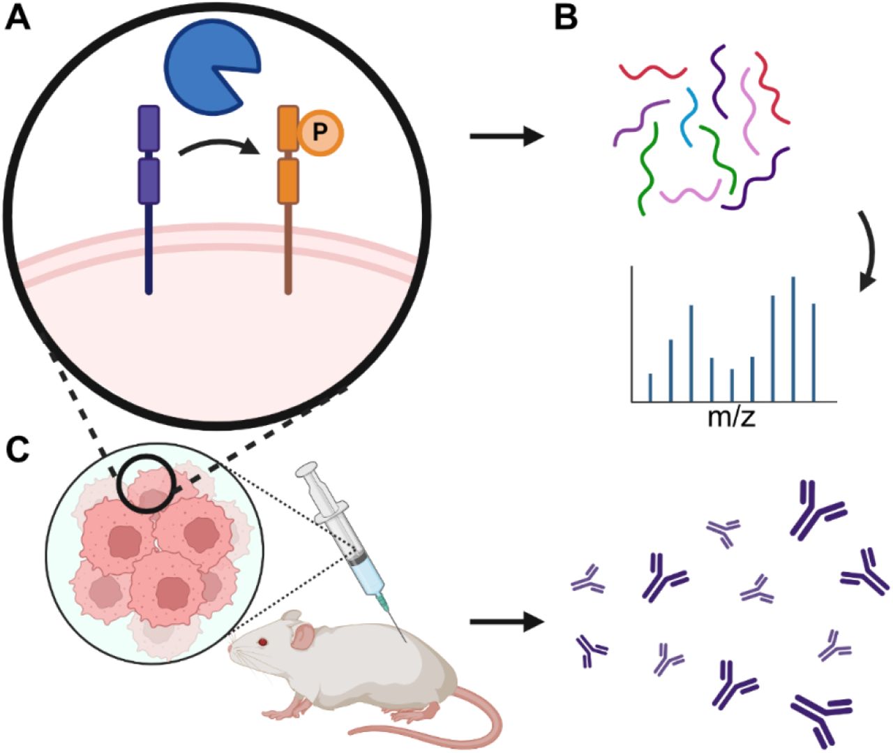
Ectokinase <t>CK2</t> has cell surface substrates that elicit an immune response. (A) Targeting kinases to cell surfaces generates phosphorylated cell surface proteins. (B) Cell surface phosphoproteins are chemically analyzed by mass spectrometry. (C) Mice immunized with whole-cell lysates of hyperphosphorylated tumor cells generate an immune response directed against cell-surface phosphorylated proteins.
Recombinant Ck2α Fusion Proteins, supplied by New England Biolabs, used in various techniques. Bioz Stars score: 86/100, based on 1 PubMed citations. ZERO BIAS - scores, article reviews, protocol conditions and more
https://www.bioz.com/product/recombinant+ck2%CE%B1/bio_rxiv__2024__03__20__585970-125-0-16?v=New+England+Biolabs
Average 86 stars, based on 1 article reviews
recombinant ck2α fusion proteins - by Bioz Stars, 2026-08
86/100 stars
  Buy from Supplier

93
Addgene inc recombinant human gst ck2α
Ectokinase <t>CK2</t> has cell surface substrates that elicit an immune response. (A) Targeting kinases to cell surfaces generates phosphorylated cell surface proteins. (B) Cell surface phosphoproteins are chemically analyzed by mass spectrometry. (C) Mice immunized with whole-cell lysates of hyperphosphorylated tumor cells generate an immune response directed against cell-surface phosphorylated proteins.
Recombinant Human Gst Ck2α, supplied by Addgene inc, used in various techniques. Bioz Stars score: 93/100, based on 1 PubMed citations. ZERO BIAS - scores, article reviews, protocol conditions and more
https://www.bioz.com/product/recombinant+ck2%CE%B1/pm38908003-168-0-4?v=Addgene+inc
Average 93 stars, based on 1 article reviews
recombinant human gst ck2α - by Bioz Stars, 2026-08
93/100 stars
  Buy from Supplier

92
OriGene recombinant ck2α protein
Fig. 5 Benzamidine, a novel <t>CK2α</t> inhibitor, reduces LRP6 stability by suppressing GRP94 phosphorylation. a CK2α kinase activity was measured after treatment with vehicle, benzamidine (10 μM and 50 μM), or silmitasertib (20 μM). Cell lysates were used for in vitro kinase assay to detect the intensity of phosphorylated substrate. Data are presented as relative activity of vehicle-treated sample. *p < 0.05; **p < 0.01 (Student’s t test). b Thermal shift assay for CK2α was performed after addition of benzamidine (10 and 50 μM). Positive derivative (d(RFU)/dT) curves are shown for control and treated with benzamidine (10 and 50 μM). Midpoint temperatures of protein-unfolding transition (Tm) are presented as bars. Values are presented as mean ± SD of at least three independent measurements. *p < 0.05; ***p < 0.001. One-way ANOVA was followed by Dunnett’s multiple comparisons test. c MDA-MB231 cells were seeded into a 12-well plate and transfected with TOPflash/ FOPflash reporter plasmids. After 24 h, cells were treated with benzamidine (10 and 50 μM) or vehicle for an additional 24 h. β-catenin transcriptional activity was assessed by measuring luminescence. TOPflash values are presented as fold change from control cells. β-galactosidase activity was used as a normalization control. Bars represent mean ± SD. *p < 0.05 (Student’s t test). d Wnt signaling-related markers were checked in MDA-MB231 cells treated with benzamidine (10 and 50 μM) for 24 h. β-actin band was used as a normalization control. e MDA-MB231 cells were transfected with Myc-GRP94 and treated with increasing concentrations of benzamidine. Cell extracts were then subjected to immunoprecipitation with anti-Myc antibody and detected with anti-p-Ser antibody. f MDA-MB231 cells were treated with benzamidine (50 μM). Stability of GRP94 and LRP6 was measured after adding CHX for the indicated time course, followed by western blotting analysis. Results are representative of at least three independent experiments and data are presented as mean ± SD.
Recombinant Ck2α Protein, supplied by OriGene, used in various techniques. Bioz Stars score: 92/100, based on 1 PubMed citations. ZERO BIAS - scores, article reviews, protocol conditions and more
https://www.bioz.com/product/recombinant+ck2%CE%B1/pm38649679-237-0-6?v=OriGene
Average 92 stars, based on 1 article reviews
recombinant ck2α protein - by Bioz Stars, 2026-08
92/100 stars
  Buy from Supplier

86
New England Biolabs recombinant human protein kinase ck2α
Protein Kinase CK2 phosphorylates of S282 and S559 . (A) 400 ng baculovirus expressed ERα was incubated in CK2 kinase buffer supplemented with 10 mM ATP, in the presence or absence of 200 ng recombinant catalytic α subunit of CK2. Reactions were stopped with Laemmli buffer, subjected to Western blot analysis, and probed with α-pS282, α-pS559, α-pS118 or αER. These studies show that the <t>CK2α</t> catalytic subunit specifically phosphorylates ERα at S282 and S559. Western blot for phosphorylation of S118, a site that exhibits strong phosphorylation in baculovirus expressed ERα, is shown for comparison to demonstrate absence of nonspecific phosphorylation by CK2 on other ERα phosphorylation sites. (B) 10 6 MCF7 breast cancer cells were pretreated with DMAT (4 uM) for 90 minutes, followed by 30 minutes with estradiol (10 -8 M) or vehicle. Immunoprecipitation of S282 or S559 was performed using phosphoantibodies and Western blot for total ERα. DMAT inhibited phosphorylation at both sites, indicating that CK2 phosphorylates these sites in vivo .
Recombinant Human Protein Kinase Ck2α, supplied by New England Biolabs, used in various techniques. Bioz Stars score: 86/100, based on 1 PubMed citations. ZERO BIAS - scores, article reviews, protocol conditions and more
https://www.bioz.com/product/recombinant+ck2%CE%B1/pmc02811108-339-15-20?v=New+England+Biolabs
Average 86 stars, based on 1 article reviews
recombinant human protein kinase ck2α - by Bioz Stars, 2026-08
86/100 stars
  Buy from Supplier

86
Thermo Fisher ck2α recombinant human protein
Protein Kinase CK2 phosphorylates of S282 and S559 . (A) 400 ng baculovirus expressed ERα was incubated in CK2 kinase buffer supplemented with 10 mM ATP, in the presence or absence of 200 ng recombinant catalytic α subunit of CK2. Reactions were stopped with Laemmli buffer, subjected to Western blot analysis, and probed with α-pS282, α-pS559, α-pS118 or αER. These studies show that the <t>CK2α</t> catalytic subunit specifically phosphorylates ERα at S282 and S559. Western blot for phosphorylation of S118, a site that exhibits strong phosphorylation in baculovirus expressed ERα, is shown for comparison to demonstrate absence of nonspecific phosphorylation by CK2 on other ERα phosphorylation sites. (B) 10 6 MCF7 breast cancer cells were pretreated with DMAT (4 uM) for 90 minutes, followed by 30 minutes with estradiol (10 -8 M) or vehicle. Immunoprecipitation of S282 or S559 was performed using phosphoantibodies and Western blot for total ERα. DMAT inhibited phosphorylation at both sites, indicating that CK2 phosphorylates these sites in vivo .
Ck2α Recombinant Human Protein, supplied by Thermo Fisher, used in various techniques. Bioz Stars score: 86/100, based on 1 PubMed citations. ZERO BIAS - scores, article reviews, protocol conditions and more
https://www.bioz.com/product/recombinant+ck2%CE%B1/pmc06148497-637-7-25?v=Thermo+Fisher
Average 86 stars, based on 1 article reviews
ck2α recombinant human protein - by Bioz Stars, 2026-08
86/100 stars
  Buy from Supplier

Image Search Results


Casein kinase 2α′ is a direct cellular target of ECH. a Identification of cellular target of ECH using pull-down technology coupled with LC–MS/MS. PC12 lysate was incubated with control beads or ECH beads. The binding proteins were detected by SDS–PAGE, followed by silver staining and LC–MS/MS analysis. b The binding proteins were detected by western blot. c Calorimetric titration of ECH with CK2α′. d ECH interacted with CK2α′, which was detected by SPR analysis. e LC–MS/MS analysis of covalently modified peptide by ECH (10 μM) in CK2α′ protein. f CK2α′ (K171A), but not CK2α′ (R173A) is responsible for ECH binding to CK2α′. HEK293T cells were transfected with HA-tagged CK2α′ and its mutant plasmids. The Lys and Arg labeled by * were potential modification sites. g Docking analysis of ECH covalent binding mode to CK2α′. h ECH increased cell viability against OGD/R-induced injury in CK2α′ −/− lung fibroblasts (LF), which were supplemented with CK2α′ but not CK2α′ mutant plasmids. i Fluorescence spectroscopy analysis of the conformational change of CK2α′ with ECH. j CD spectra analysis for ECH-mediated CK2α′ conformational change. Data are expressed as the mean ± SD. ## P < 0.01 vs. control group, * P < 0.05, ** P < 0.01 vs. OGD/R group

Journal: Signal Transduction and Targeted Therapy

Article Title: Small molecule induces mitochondrial fusion for neuroprotection via targeting CK2 without affecting its conventional kinase activity

doi: 10.1038/s41392-020-00447-6

Figure Lengend Snippet: Casein kinase 2α′ is a direct cellular target of ECH. a Identification of cellular target of ECH using pull-down technology coupled with LC–MS/MS. PC12 lysate was incubated with control beads or ECH beads. The binding proteins were detected by SDS–PAGE, followed by silver staining and LC–MS/MS analysis. b The binding proteins were detected by western blot. c Calorimetric titration of ECH with CK2α′. d ECH interacted with CK2α′, which was detected by SPR analysis. e LC–MS/MS analysis of covalently modified peptide by ECH (10 μM) in CK2α′ protein. f CK2α′ (K171A), but not CK2α′ (R173A) is responsible for ECH binding to CK2α′. HEK293T cells were transfected with HA-tagged CK2α′ and its mutant plasmids. The Lys and Arg labeled by * were potential modification sites. g Docking analysis of ECH covalent binding mode to CK2α′. h ECH increased cell viability against OGD/R-induced injury in CK2α′ −/− lung fibroblasts (LF), which were supplemented with CK2α′ but not CK2α′ mutant plasmids. i Fluorescence spectroscopy analysis of the conformational change of CK2α′ with ECH. j CD spectra analysis for ECH-mediated CK2α′ conformational change. Data are expressed as the mean ± SD. ## P < 0.01 vs. control group, * P < 0.05, ** P < 0.01 vs. OGD/R group

Article Snippet: Human recombinant CK2α′ protein was purchased from Sino Biological (Beijing, China).

Techniques: Liquid Chromatography with Mass Spectroscopy, Incubation, Binding Assay, SDS Page, Silver Staining, Western Blot, Titration, Modification, Transfection, Mutagenesis, Labeling, Fluorescence, Spectroscopy

CK2α′ plays a fundamental role in modulating mitochondrial fusion. a CK2α′ knock-down reversed ECH-dependent increase in cell viability against OGD/R insult, which was detected by MTT assay. b ECH increased cell viability against OGD/R-induced injury in CK2α′ +/+ lung fibroblasts, which was significantly suppressed in CK2α′ −/− cells. c ECH-induced mitochondrial fusion was suppressed in CK2α′ knock-down cells, which was indicated by Mito-tracker staining. Arrows (red) indicate branched healthy mitochondria. Arrows (white) indicate spherical dysfunctional mitochondria (scale bar: 20 μm). d ECH-induced mitochondrial fusion was inhibited in CK2α′ −/− cells, which was indicated by Mito-tracker staining. Arrows (red) indicate branched healthy mitochondria. Arrows (white) indicate spherical dysfunctional mitochondria (scale bar: 20 μm). e ECH recovered mitochondrial fusion induction in CK2α′ −/− lung fibroblasts which were transfected with CK2α′ plasmid. Arrows (red) indicate branched healthy mitochondria. Arrows (white) indicate spherical dysfunctional mitochondria (scale bar: 20 μm). f Mfn2 expression induced by ECH was downregulated in CK2α′ knock-down PC12 cells, which was analyzed by western blot. Data are expressed as the mean ± SD. ## P < 0.01 vs. control group, * P < 0.05, ** P < 0.01 vs. OGD/R group. NS not significant

Journal: Signal Transduction and Targeted Therapy

Article Title: Small molecule induces mitochondrial fusion for neuroprotection via targeting CK2 without affecting its conventional kinase activity

doi: 10.1038/s41392-020-00447-6

Figure Lengend Snippet: CK2α′ plays a fundamental role in modulating mitochondrial fusion. a CK2α′ knock-down reversed ECH-dependent increase in cell viability against OGD/R insult, which was detected by MTT assay. b ECH increased cell viability against OGD/R-induced injury in CK2α′ +/+ lung fibroblasts, which was significantly suppressed in CK2α′ −/− cells. c ECH-induced mitochondrial fusion was suppressed in CK2α′ knock-down cells, which was indicated by Mito-tracker staining. Arrows (red) indicate branched healthy mitochondria. Arrows (white) indicate spherical dysfunctional mitochondria (scale bar: 20 μm). d ECH-induced mitochondrial fusion was inhibited in CK2α′ −/− cells, which was indicated by Mito-tracker staining. Arrows (red) indicate branched healthy mitochondria. Arrows (white) indicate spherical dysfunctional mitochondria (scale bar: 20 μm). e ECH recovered mitochondrial fusion induction in CK2α′ −/− lung fibroblasts which were transfected with CK2α′ plasmid. Arrows (red) indicate branched healthy mitochondria. Arrows (white) indicate spherical dysfunctional mitochondria (scale bar: 20 μm). f Mfn2 expression induced by ECH was downregulated in CK2α′ knock-down PC12 cells, which was analyzed by western blot. Data are expressed as the mean ± SD. ## P < 0.01 vs. control group, * P < 0.05, ** P < 0.01 vs. OGD/R group. NS not significant

Article Snippet: Human recombinant CK2α′ protein was purchased from Sino Biological (Beijing, China).

Techniques: MTT Assay, Staining, Transfection, Plasmid Preparation, Expressing, Western Blot

CK2α′ binds to basic transcription factor (BTF3) as a direct substrate. a Stable isotope labeling with amino acids in cell culture (SILAC)-coupled pull-down technology was used to identify the binding substrates of CK2α′. b Overview of the binding substrates of CK2α′ was identified by LC–MS/MS. c BTF3 knock-down inhibited ECH-mediated cell viability increase, which was detected by MTT assay. d ECH-induced mitochondrial fusion was inhibited in BTF3 knock-down cells, which was indicated by Mito-tracker staining. Arrows (red) indicate branched healthy mitochondria. Arrows (white) indicate spherical dysfunctional mitochondria (scale bar: 20 μm). e BTF3 knock-down inhibited Mfn2 expression, which was detected by western blot. f Genome browser view of BTF3α ChIP-seq signal on Mfn2 gene loci. g Top enriched de novo transcription factor (TF) motifs within 14 bp upon ECH treatment. h Co-localization of β-catenin (red) and DAPI (blue) was detected by immunofluorescent staining. Arrows (red) indicate cytoplasmic location of β-catenin. Arrows (white) indicate nuclear translocation of β-catenin from the cytoplasm (scale bar: 20 μm). i ECH facilitated β-catenin nuclear translocation. j IWP-2 reversed ECH-dependent cell viability increase under OGD/R insult. Data are expressed as the mean ± SD. ## P < 0.01 vs. control group, * P < 0.05, ** P < 0.01 vs. OGD/R group

Journal: Signal Transduction and Targeted Therapy

Article Title: Small molecule induces mitochondrial fusion for neuroprotection via targeting CK2 without affecting its conventional kinase activity

doi: 10.1038/s41392-020-00447-6

Figure Lengend Snippet: CK2α′ binds to basic transcription factor (BTF3) as a direct substrate. a Stable isotope labeling with amino acids in cell culture (SILAC)-coupled pull-down technology was used to identify the binding substrates of CK2α′. b Overview of the binding substrates of CK2α′ was identified by LC–MS/MS. c BTF3 knock-down inhibited ECH-mediated cell viability increase, which was detected by MTT assay. d ECH-induced mitochondrial fusion was inhibited in BTF3 knock-down cells, which was indicated by Mito-tracker staining. Arrows (red) indicate branched healthy mitochondria. Arrows (white) indicate spherical dysfunctional mitochondria (scale bar: 20 μm). e BTF3 knock-down inhibited Mfn2 expression, which was detected by western blot. f Genome browser view of BTF3α ChIP-seq signal on Mfn2 gene loci. g Top enriched de novo transcription factor (TF) motifs within 14 bp upon ECH treatment. h Co-localization of β-catenin (red) and DAPI (blue) was detected by immunofluorescent staining. Arrows (red) indicate cytoplasmic location of β-catenin. Arrows (white) indicate nuclear translocation of β-catenin from the cytoplasm (scale bar: 20 μm). i ECH facilitated β-catenin nuclear translocation. j IWP-2 reversed ECH-dependent cell viability increase under OGD/R insult. Data are expressed as the mean ± SD. ## P < 0.01 vs. control group, * P < 0.05, ** P < 0.01 vs. OGD/R group

Article Snippet: Human recombinant CK2α′ protein was purchased from Sino Biological (Beijing, China).

Techniques: Labeling, Cell Culture, Binding Assay, Liquid Chromatography with Mass Spectroscopy, MTT Assay, Staining, Expressing, Western Blot, ChIP-sequencing, Translocation Assay

BTF3 promotes Mfn2 expression in Wnt/β-catenin signal-dependent manner. a , b ECH enhanced TCF/LEF1 reporter gene activity, which was inhibited by CK2α′ or BTF3 knock-down. c ECH inhibited the interaction of BTF3α with TCF1/7 and LEF1. HEK293T cells were transfected with HA-tagged BTF3α and treated by ECH (10 μM) for 6 h. Co-IP was performed with anti-HA antibody, followed by immunoblotting analysis. d Cytoplasmic translocation of HA-BTF3α (green) from nucleus and nuclear translocation of β-catenin (red) from cytoplasm were promoted by ECH (scale bar: 20 μm). e IWP-2 blocked ECH-induced mitochondrial fusion; however, TWS119 did not show similar effect. Arrows (red) indicate branched healthy mitochondria. Arrows (white) indicate spherical dysfunctional mitochondria (scale bar: 20 μm). Data are expressed as the mean ± SD. ## P < 0.01 vs. control group. * P < 0.05, ** P < 0.01 vs. OGD/R group

Journal: Signal Transduction and Targeted Therapy

Article Title: Small molecule induces mitochondrial fusion for neuroprotection via targeting CK2 without affecting its conventional kinase activity

doi: 10.1038/s41392-020-00447-6

Figure Lengend Snippet: BTF3 promotes Mfn2 expression in Wnt/β-catenin signal-dependent manner. a , b ECH enhanced TCF/LEF1 reporter gene activity, which was inhibited by CK2α′ or BTF3 knock-down. c ECH inhibited the interaction of BTF3α with TCF1/7 and LEF1. HEK293T cells were transfected with HA-tagged BTF3α and treated by ECH (10 μM) for 6 h. Co-IP was performed with anti-HA antibody, followed by immunoblotting analysis. d Cytoplasmic translocation of HA-BTF3α (green) from nucleus and nuclear translocation of β-catenin (red) from cytoplasm were promoted by ECH (scale bar: 20 μm). e IWP-2 blocked ECH-induced mitochondrial fusion; however, TWS119 did not show similar effect. Arrows (red) indicate branched healthy mitochondria. Arrows (white) indicate spherical dysfunctional mitochondria (scale bar: 20 μm). Data are expressed as the mean ± SD. ## P < 0.01 vs. control group. * P < 0.05, ** P < 0.01 vs. OGD/R group

Article Snippet: Human recombinant CK2α′ protein was purchased from Sino Biological (Beijing, China).

Techniques: Expressing, Activity Assay, Transfection, Co-Immunoprecipitation Assay, Western Blot, Translocation Assay

CK2 exerts neuroprotection via promoting mitochondrial fusion in MCAO model. a Scheme of neuroprotection evaluation of ECH in MCAO model. MCAO were established on mice and ECH (50 mg/kg) was administrated for every 2 days, followed by neurological score test in day 9 and bioassay in day 10. b ECH improved neurological score against MCAO insult in CK2α′ +/+ mice, but showed no effect in CK2α′ +/− mice. c ECH protected neurons against MCAO insult in CK2α′ +/+ mice, but showed no effect in CK2α′ +/− mice. Neuroprotection in hippocampal and cortical areas were detected by Nissl staining assay. Arrows indicate Nissl bodies (scale bar: 50 μm). d ECH inhibited cell apoptosis against MCAO insult in CK2α′ +/+ mice, but showed no effect in CK2α′ +/− mice. Cell apoptosis was detected by TUNEL assay (scale bar: 50 µm). e Mfn2 expression was increased by ECH in CK2α′ +/+ mice, but not in CK2α′ +/− mice (scale bar: 50 μm). f ECH-induced mitochondrial fusion was suppressed in CK2α′ +/− mice, which was stained with the specific mitochondrial marker COXIV (scale bar: 50 μm). g CK2α′ +/− blocked ECH-induced Mfn2 expression increase, which was detected by western blot assay. h TCF1/7 significantly translocated into nucleus upon ECH treatment (scale bar: 50 μm). i , j Swimming performance analysis for ECH and IWP-2 neuroprotection in ischemia/reperfusion-induced zebrafishes. Data are expressed as the mean ± SD. ## P < 0.01 vs. control group. * P < 0.05, ** P < 0.01 vs. MCAO group. NS not significant

Journal: Signal Transduction and Targeted Therapy

Article Title: Small molecule induces mitochondrial fusion for neuroprotection via targeting CK2 without affecting its conventional kinase activity

doi: 10.1038/s41392-020-00447-6

Figure Lengend Snippet: CK2 exerts neuroprotection via promoting mitochondrial fusion in MCAO model. a Scheme of neuroprotection evaluation of ECH in MCAO model. MCAO were established on mice and ECH (50 mg/kg) was administrated for every 2 days, followed by neurological score test in day 9 and bioassay in day 10. b ECH improved neurological score against MCAO insult in CK2α′ +/+ mice, but showed no effect in CK2α′ +/− mice. c ECH protected neurons against MCAO insult in CK2α′ +/+ mice, but showed no effect in CK2α′ +/− mice. Neuroprotection in hippocampal and cortical areas were detected by Nissl staining assay. Arrows indicate Nissl bodies (scale bar: 50 μm). d ECH inhibited cell apoptosis against MCAO insult in CK2α′ +/+ mice, but showed no effect in CK2α′ +/− mice. Cell apoptosis was detected by TUNEL assay (scale bar: 50 µm). e Mfn2 expression was increased by ECH in CK2α′ +/+ mice, but not in CK2α′ +/− mice (scale bar: 50 μm). f ECH-induced mitochondrial fusion was suppressed in CK2α′ +/− mice, which was stained with the specific mitochondrial marker COXIV (scale bar: 50 μm). g CK2α′ +/− blocked ECH-induced Mfn2 expression increase, which was detected by western blot assay. h TCF1/7 significantly translocated into nucleus upon ECH treatment (scale bar: 50 μm). i , j Swimming performance analysis for ECH and IWP-2 neuroprotection in ischemia/reperfusion-induced zebrafishes. Data are expressed as the mean ± SD. ## P < 0.01 vs. control group. * P < 0.05, ** P < 0.01 vs. MCAO group. NS not significant

Article Snippet: Human recombinant CK2α′ protein was purchased from Sino Biological (Beijing, China).

Techniques: Staining, TUNEL Assay, Expressing, Marker, Western Blot

Ectokinase CK2 has cell surface substrates that elicit an immune response. (A) Targeting kinases to cell surfaces generates phosphorylated cell surface proteins. (B) Cell surface phosphoproteins are chemically analyzed by mass spectrometry. (C) Mice immunized with whole-cell lysates of hyperphosphorylated tumor cells generate an immune response directed against cell-surface phosphorylated proteins.

Journal: bioRxiv

Article Title: Chemoproteomics reveals immunogenic and tumor-associated cell surface substrates of ectokinase CK2α

doi: 10.1101/2024.03.20.585970

Figure Lengend Snippet: Ectokinase CK2 has cell surface substrates that elicit an immune response. (A) Targeting kinases to cell surfaces generates phosphorylated cell surface proteins. (B) Cell surface phosphoproteins are chemically analyzed by mass spectrometry. (C) Mice immunized with whole-cell lysates of hyperphosphorylated tumor cells generate an immune response directed against cell-surface phosphorylated proteins.

Article Snippet: Recombinant CK2α fusion proteins were expressed as maltose binding protein (MBP) conjugates in BL21(DE3) E. coli (NEB, C2527H) via autoinduction.

Techniques: Mass Spectrometry

Cell-tethered CK2α is active on the cell surface under tumor-relevant conditions. (A) Schematic representation of kinase-fusion proteins generated. (B) Purified recombinant ZHER2-CK2α or CK2α proteins, C-terminally fused to a cMyc peptide tag, were incubated with hHER2-expressing EMT-6 murine breast cancer cells, and kinase binding was visualized with a fluorophore-conjugated anti-cMyc antibody by flow cytometry. (C) Generation of CK2α isoforms that are more stable and active. CK2α mutants were generated and assayed for activity on a model peptide substrate (RRRDDDSDDD) in tumor-relevant concentrations of ATP (100 micromolar). (D,E) hHER2 + EMT-6 cells were incubated with kinase fusion and ATP-gamma-thiophosphate in kinase reaction buffer, before being lysed, alkylated with p-nitrobenzylmesylate, separated by SDS-PAGE, and transferred to a blotting membrane. Ectophosphorylation was visualized with a thioester-specific antibody and quantitated by near-infrared fluorescence imaging. ( D ) Representative Western Blot. ( E ) Fluorescence quantitation from three independent blots. Statistics were determined by one-way ANOVA corrected for multiple comparisons. * = corrected p value < 0.05. n.s. = not significant. Corrected p-values are displayed above relevant comparisons.

Journal: bioRxiv

Article Title: Chemoproteomics reveals immunogenic and tumor-associated cell surface substrates of ectokinase CK2α

doi: 10.1101/2024.03.20.585970

Figure Lengend Snippet: Cell-tethered CK2α is active on the cell surface under tumor-relevant conditions. (A) Schematic representation of kinase-fusion proteins generated. (B) Purified recombinant ZHER2-CK2α or CK2α proteins, C-terminally fused to a cMyc peptide tag, were incubated with hHER2-expressing EMT-6 murine breast cancer cells, and kinase binding was visualized with a fluorophore-conjugated anti-cMyc antibody by flow cytometry. (C) Generation of CK2α isoforms that are more stable and active. CK2α mutants were generated and assayed for activity on a model peptide substrate (RRRDDDSDDD) in tumor-relevant concentrations of ATP (100 micromolar). (D,E) hHER2 + EMT-6 cells were incubated with kinase fusion and ATP-gamma-thiophosphate in kinase reaction buffer, before being lysed, alkylated with p-nitrobenzylmesylate, separated by SDS-PAGE, and transferred to a blotting membrane. Ectophosphorylation was visualized with a thioester-specific antibody and quantitated by near-infrared fluorescence imaging. ( D ) Representative Western Blot. ( E ) Fluorescence quantitation from three independent blots. Statistics were determined by one-way ANOVA corrected for multiple comparisons. * = corrected p value < 0.05. n.s. = not significant. Corrected p-values are displayed above relevant comparisons.

Article Snippet: Recombinant CK2α fusion proteins were expressed as maltose binding protein (MBP) conjugates in BL21(DE3) E. coli (NEB, C2527H) via autoinduction.

Techniques: Generated, Purification, Recombinant, Incubation, Expressing, Binding Assay, Flow Cytometry, Activity Assay, SDS Page, Membrane, Fluorescence, Imaging, Western Blot, Quantitation Assay

Identification of CK2 substrates using an ATP-gamma-thiophosphate (ATPγS) chemical reporter substrate using label-free quantitation based proteomics. (A,B) Wild-type or loss-of-function CK2α is tethered to the cell surface in the presence of ATPγS, lysed, immobilized on iodoacetyl agarose, and then tryptic eluates are analyzed by LC-MS (B). (C) Candidate recombinant CK2 substrates were treated in vitro with recombinant CK2, alkylated with p-nitrobenzylmesylate, and analyzed by Western blot with a thiophosphate ester-specific antibody. (D) Wild-type or loss-of-function CK2α was tethered to the surface of hHER2+ EMT-6 cells in the presence of ATPγS. Cells were lysed and CD44 was immunoprecipitated from lysates. Immunopreciptates were alkylated with p-nitrobenzylmesylate and analyzed by Western blot with a thiophosphate ester-specific antibody showing active kinase-dependent increase in CD44 phosphorylation.

Journal: bioRxiv

Article Title: Chemoproteomics reveals immunogenic and tumor-associated cell surface substrates of ectokinase CK2α

doi: 10.1101/2024.03.20.585970

Figure Lengend Snippet: Identification of CK2 substrates using an ATP-gamma-thiophosphate (ATPγS) chemical reporter substrate using label-free quantitation based proteomics. (A,B) Wild-type or loss-of-function CK2α is tethered to the cell surface in the presence of ATPγS, lysed, immobilized on iodoacetyl agarose, and then tryptic eluates are analyzed by LC-MS (B). (C) Candidate recombinant CK2 substrates were treated in vitro with recombinant CK2, alkylated with p-nitrobenzylmesylate, and analyzed by Western blot with a thiophosphate ester-specific antibody. (D) Wild-type or loss-of-function CK2α was tethered to the surface of hHER2+ EMT-6 cells in the presence of ATPγS. Cells were lysed and CD44 was immunoprecipitated from lysates. Immunopreciptates were alkylated with p-nitrobenzylmesylate and analyzed by Western blot with a thiophosphate ester-specific antibody showing active kinase-dependent increase in CD44 phosphorylation.

Article Snippet: Recombinant CK2α fusion proteins were expressed as maltose binding protein (MBP) conjugates in BL21(DE3) E. coli (NEB, C2527H) via autoinduction.

Techniques: Quantitation Assay, Liquid Chromatography with Mass Spectroscopy, Recombinant, In Vitro, Western Blot, Immunoprecipitation

Whole-cell vaccination with hyperphosphorylated cell surfaces elicits a humoral but not a tumor-killing immune response. (A) Mice were immunized weekly for four weeks with heat-killed EMT-6 cells that had either been hyperphosphorylated with cell-tethered CK2α in vitro or treated with vehicle. Serum was collected and measured for binding to syngenic tumor cells or mice were challenged with live tumor cell implantation. (B) Binding of sera from five mice immunized with hyperphosphorylated EMT-6 cells (Group 1) or control cells (Group 2) to live EMT-6 pretreated with cell-tethered wild-type (CK2α -treated) or loss-of-function (control treated) CK2α in vitro shows significant increase in antibodies for cells treated with wild-type CK2α. Binding of mouse IgG was visualized by the binding of a rabbit anti-mIgG AlexaFluor647 conjugate. (C) Binding of immunized sera from Group 1 or Group 2 to EMT-6 cells pretreated with N6-phenylethyl ATP or vehicle and CK2α F113G or vehicle. Statistics for (B) and (C) were determined by corrected multiple t-tests. (D) Mice were then challenged with live EMT-6 cells that were either hyperphosphorylated with cell-tethered CK2α or treated with vehicle in vitro prior to implantation. Averaged tumor growth curves from mice immunized with hyperphosphorylated EMT-6 cells. For ( D ) error bars represent standard deviation of 5 mice per arm. * = corrected p value < 0.05. ** = corrected p value < 0.01. Corrected p-values are displayed above relevant comparisons. Non-significant comparisons are not displayed.

Journal: bioRxiv

Article Title: Chemoproteomics reveals immunogenic and tumor-associated cell surface substrates of ectokinase CK2α

doi: 10.1101/2024.03.20.585970

Figure Lengend Snippet: Whole-cell vaccination with hyperphosphorylated cell surfaces elicits a humoral but not a tumor-killing immune response. (A) Mice were immunized weekly for four weeks with heat-killed EMT-6 cells that had either been hyperphosphorylated with cell-tethered CK2α in vitro or treated with vehicle. Serum was collected and measured for binding to syngenic tumor cells or mice were challenged with live tumor cell implantation. (B) Binding of sera from five mice immunized with hyperphosphorylated EMT-6 cells (Group 1) or control cells (Group 2) to live EMT-6 pretreated with cell-tethered wild-type (CK2α -treated) or loss-of-function (control treated) CK2α in vitro shows significant increase in antibodies for cells treated with wild-type CK2α. Binding of mouse IgG was visualized by the binding of a rabbit anti-mIgG AlexaFluor647 conjugate. (C) Binding of immunized sera from Group 1 or Group 2 to EMT-6 cells pretreated with N6-phenylethyl ATP or vehicle and CK2α F113G or vehicle. Statistics for (B) and (C) were determined by corrected multiple t-tests. (D) Mice were then challenged with live EMT-6 cells that were either hyperphosphorylated with cell-tethered CK2α or treated with vehicle in vitro prior to implantation. Averaged tumor growth curves from mice immunized with hyperphosphorylated EMT-6 cells. For ( D ) error bars represent standard deviation of 5 mice per arm. * = corrected p value < 0.05. ** = corrected p value < 0.01. Corrected p-values are displayed above relevant comparisons. Non-significant comparisons are not displayed.

Article Snippet: Recombinant CK2α fusion proteins were expressed as maltose binding protein (MBP) conjugates in BL21(DE3) E. coli (NEB, C2527H) via autoinduction.

Techniques: In Vitro, Binding Assay, Standard Deviation

Immunization with CK2 surface substrates elicits a humoral immune response . (A-D) Immunogens were identified by binding sera from either Group 1 (wild-type ZHER2-CK2α) or Group 2 (inactive ZHER2-CK2α) mice to hHER2+ EMT-6 pretreated with cell-tethered CK2α, gently lysing the cells, precipitating mIgG on immobilized Protein A, and analyzing tryptic digests by LC-MS. (B) Identification of unique proteins from -immunized sera shows many more for the wild-type ZHER2-CK2α treated mice. (C) rmCD44 was phosphorylated in vitro with CK2 and tested for binding sera by ELISA shows positive sera for the wild-type ZHER2-CK2α treated mice. (D) VH genes were subcloned from splenocyte mRNA and ported into a scFv-based yeast-display vector for enrichment of phospho-CD44 binding clones. Top 5 enriched clones were expressed with a TwinStrep affinity tag and binding to hHER2+ EMT-6 cells pretreated with cell-tethered CK2α was quantified using a fluorescent Streptactin-XT. Statistics were determined by multiple t-tests corrected for multiple comparisons by the Holm-Sidak method. (E-G) Splenocytes from immunized mice were characterized by flow cytometry. (E) Germinal B cells were quantitated as a proportion of total B cells. Statistics determined by Student’s T test. (F,G) Splenocytes were stimulated with either antibody cocktails or hHER2+ EMT-6 cell, pretreated with cell-tethered CK2α, lysates and allowed to activate for 72 hours. Analysis of the activation state of CD8+ T cells (F) and CD4+ T cells (G) was measured by upregulation of CD25 by flow cytometry. Statistics for (F,G) were determined by corrected multiple T tests. * = corrected p value < 0.05. Corrected p-values are displayed above relevant comparisons. Non-significant comparisons are not displayed.

Journal: bioRxiv

Article Title: Chemoproteomics reveals immunogenic and tumor-associated cell surface substrates of ectokinase CK2α

doi: 10.1101/2024.03.20.585970

Figure Lengend Snippet: Immunization with CK2 surface substrates elicits a humoral immune response . (A-D) Immunogens were identified by binding sera from either Group 1 (wild-type ZHER2-CK2α) or Group 2 (inactive ZHER2-CK2α) mice to hHER2+ EMT-6 pretreated with cell-tethered CK2α, gently lysing the cells, precipitating mIgG on immobilized Protein A, and analyzing tryptic digests by LC-MS. (B) Identification of unique proteins from -immunized sera shows many more for the wild-type ZHER2-CK2α treated mice. (C) rmCD44 was phosphorylated in vitro with CK2 and tested for binding sera by ELISA shows positive sera for the wild-type ZHER2-CK2α treated mice. (D) VH genes were subcloned from splenocyte mRNA and ported into a scFv-based yeast-display vector for enrichment of phospho-CD44 binding clones. Top 5 enriched clones were expressed with a TwinStrep affinity tag and binding to hHER2+ EMT-6 cells pretreated with cell-tethered CK2α was quantified using a fluorescent Streptactin-XT. Statistics were determined by multiple t-tests corrected for multiple comparisons by the Holm-Sidak method. (E-G) Splenocytes from immunized mice were characterized by flow cytometry. (E) Germinal B cells were quantitated as a proportion of total B cells. Statistics determined by Student’s T test. (F,G) Splenocytes were stimulated with either antibody cocktails or hHER2+ EMT-6 cell, pretreated with cell-tethered CK2α, lysates and allowed to activate for 72 hours. Analysis of the activation state of CD8+ T cells (F) and CD4+ T cells (G) was measured by upregulation of CD25 by flow cytometry. Statistics for (F,G) were determined by corrected multiple T tests. * = corrected p value < 0.05. Corrected p-values are displayed above relevant comparisons. Non-significant comparisons are not displayed.

Article Snippet: Recombinant CK2α fusion proteins were expressed as maltose binding protein (MBP) conjugates in BL21(DE3) E. coli (NEB, C2527H) via autoinduction.

Techniques: Binding Assay, Liquid Chromatography with Mass Spectroscopy, In Vitro, Enzyme-linked Immunosorbent Assay, Plasmid Preparation, Clone Assay, Flow Cytometry, Activation Assay

Fig. 5 Benzamidine, a novel CK2α inhibitor, reduces LRP6 stability by suppressing GRP94 phosphorylation. a CK2α kinase activity was measured after treatment with vehicle, benzamidine (10 μM and 50 μM), or silmitasertib (20 μM). Cell lysates were used for in vitro kinase assay to detect the intensity of phosphorylated substrate. Data are presented as relative activity of vehicle-treated sample. *p < 0.05; **p < 0.01 (Student’s t test). b Thermal shift assay for CK2α was performed after addition of benzamidine (10 and 50 μM). Positive derivative (d(RFU)/dT) curves are shown for control and treated with benzamidine (10 and 50 μM). Midpoint temperatures of protein-unfolding transition (Tm) are presented as bars. Values are presented as mean ± SD of at least three independent measurements. *p < 0.05; ***p < 0.001. One-way ANOVA was followed by Dunnett’s multiple comparisons test. c MDA-MB231 cells were seeded into a 12-well plate and transfected with TOPflash/ FOPflash reporter plasmids. After 24 h, cells were treated with benzamidine (10 and 50 μM) or vehicle for an additional 24 h. β-catenin transcriptional activity was assessed by measuring luminescence. TOPflash values are presented as fold change from control cells. β-galactosidase activity was used as a normalization control. Bars represent mean ± SD. *p < 0.05 (Student’s t test). d Wnt signaling-related markers were checked in MDA-MB231 cells treated with benzamidine (10 and 50 μM) for 24 h. β-actin band was used as a normalization control. e MDA-MB231 cells were transfected with Myc-GRP94 and treated with increasing concentrations of benzamidine. Cell extracts were then subjected to immunoprecipitation with anti-Myc antibody and detected with anti-p-Ser antibody. f MDA-MB231 cells were treated with benzamidine (50 μM). Stability of GRP94 and LRP6 was measured after adding CHX for the indicated time course, followed by western blotting analysis. Results are representative of at least three independent experiments and data are presented as mean ± SD.

Journal: Cell death discovery

Article Title: CK2α-mediated phosphorylation of GRP94 facilitates the metastatic cascade in triple-negative breast cancer.

doi: 10.1038/s41420-024-01956-x

Figure Lengend Snippet: Fig. 5 Benzamidine, a novel CK2α inhibitor, reduces LRP6 stability by suppressing GRP94 phosphorylation. a CK2α kinase activity was measured after treatment with vehicle, benzamidine (10 μM and 50 μM), or silmitasertib (20 μM). Cell lysates were used for in vitro kinase assay to detect the intensity of phosphorylated substrate. Data are presented as relative activity of vehicle-treated sample. *p < 0.05; **p < 0.01 (Student’s t test). b Thermal shift assay for CK2α was performed after addition of benzamidine (10 and 50 μM). Positive derivative (d(RFU)/dT) curves are shown for control and treated with benzamidine (10 and 50 μM). Midpoint temperatures of protein-unfolding transition (Tm) are presented as bars. Values are presented as mean ± SD of at least three independent measurements. *p < 0.05; ***p < 0.001. One-way ANOVA was followed by Dunnett’s multiple comparisons test. c MDA-MB231 cells were seeded into a 12-well plate and transfected with TOPflash/ FOPflash reporter plasmids. After 24 h, cells were treated with benzamidine (10 and 50 μM) or vehicle for an additional 24 h. β-catenin transcriptional activity was assessed by measuring luminescence. TOPflash values are presented as fold change from control cells. β-galactosidase activity was used as a normalization control. Bars represent mean ± SD. *p < 0.05 (Student’s t test). d Wnt signaling-related markers were checked in MDA-MB231 cells treated with benzamidine (10 and 50 μM) for 24 h. β-actin band was used as a normalization control. e MDA-MB231 cells were transfected with Myc-GRP94 and treated with increasing concentrations of benzamidine. Cell extracts were then subjected to immunoprecipitation with anti-Myc antibody and detected with anti-p-Ser antibody. f MDA-MB231 cells were treated with benzamidine (50 μM). Stability of GRP94 and LRP6 was measured after adding CHX for the indicated time course, followed by western blotting analysis. Results are representative of at least three independent experiments and data are presented as mean ± SD.

Article Snippet: Recombinant CK2α protein was obtained from Origene (TP760181, Rockville, MD, USA).

Techniques: Phospho-proteomics, Activity Assay, In Vitro, Kinase Assay, Thermal Shift Assay, Control, Transfection, Immunoprecipitation, Western Blot

Fig. 7 Schematic diagram illustrating the important role of CK2α- GRP94-LRP6 axis in TNBC metastasis. In TNBC cells, elevated CK2α phosphorylates GRP94 at serine 306 residue, leading to increased stability of the GRP94 protein. As an ER chaperone, GRP94 promotes the maturation of LRP6 protein, resulting in activation of Wnt signaling. Collectively, the activation and upregulation of CK2α and GRP94 synergistically promote the metastasis of TNBC cells through the Wnt signaling pathway. By targeting CK2α-mediated phosphor- ylation of GRP94 with benzamidine, metastasis of TNBC can be effectively controlled as a therapeutic strategy.

Journal: Cell death discovery

Article Title: CK2α-mediated phosphorylation of GRP94 facilitates the metastatic cascade in triple-negative breast cancer.

doi: 10.1038/s41420-024-01956-x

Figure Lengend Snippet: Fig. 7 Schematic diagram illustrating the important role of CK2α- GRP94-LRP6 axis in TNBC metastasis. In TNBC cells, elevated CK2α phosphorylates GRP94 at serine 306 residue, leading to increased stability of the GRP94 protein. As an ER chaperone, GRP94 promotes the maturation of LRP6 protein, resulting in activation of Wnt signaling. Collectively, the activation and upregulation of CK2α and GRP94 synergistically promote the metastasis of TNBC cells through the Wnt signaling pathway. By targeting CK2α-mediated phosphor- ylation of GRP94 with benzamidine, metastasis of TNBC can be effectively controlled as a therapeutic strategy.

Article Snippet: Recombinant CK2α protein was obtained from Origene (TP760181, Rockville, MD, USA).

Techniques: Residue, Activation Assay

Protein Kinase CK2 phosphorylates of S282 and S559 . (A) 400 ng baculovirus expressed ERα was incubated in CK2 kinase buffer supplemented with 10 mM ATP, in the presence or absence of 200 ng recombinant catalytic α subunit of CK2. Reactions were stopped with Laemmli buffer, subjected to Western blot analysis, and probed with α-pS282, α-pS559, α-pS118 or αER. These studies show that the CK2α catalytic subunit specifically phosphorylates ERα at S282 and S559. Western blot for phosphorylation of S118, a site that exhibits strong phosphorylation in baculovirus expressed ERα, is shown for comparison to demonstrate absence of nonspecific phosphorylation by CK2 on other ERα phosphorylation sites. (B) 10 6 MCF7 breast cancer cells were pretreated with DMAT (4 uM) for 90 minutes, followed by 30 minutes with estradiol (10 -8 M) or vehicle. Immunoprecipitation of S282 or S559 was performed using phosphoantibodies and Western blot for total ERα. DMAT inhibited phosphorylation at both sites, indicating that CK2 phosphorylates these sites in vivo .

Journal: BMC Biochemistry

Article Title: Identification of four novel phosphorylation sites in estrogen receptor α: impact on receptor-dependent gene expression and phosphorylation by protein kinase CK2

doi: 10.1186/1471-2091-10-36

Figure Lengend Snippet: Protein Kinase CK2 phosphorylates of S282 and S559 . (A) 400 ng baculovirus expressed ERα was incubated in CK2 kinase buffer supplemented with 10 mM ATP, in the presence or absence of 200 ng recombinant catalytic α subunit of CK2. Reactions were stopped with Laemmli buffer, subjected to Western blot analysis, and probed with α-pS282, α-pS559, α-pS118 or αER. These studies show that the CK2α catalytic subunit specifically phosphorylates ERα at S282 and S559. Western blot for phosphorylation of S118, a site that exhibits strong phosphorylation in baculovirus expressed ERα, is shown for comparison to demonstrate absence of nonspecific phosphorylation by CK2 on other ERα phosphorylation sites. (B) 10 6 MCF7 breast cancer cells were pretreated with DMAT (4 uM) for 90 minutes, followed by 30 minutes with estradiol (10 -8 M) or vehicle. Immunoprecipitation of S282 or S559 was performed using phosphoantibodies and Western blot for total ERα. DMAT inhibited phosphorylation at both sites, indicating that CK2 phosphorylates these sites in vivo .

Article Snippet: Briefly, purified baculovirus expressed ERα (300 ng), (Invitrogen, Carlsbad, CA) was incubated with 100 ng recombinant human protein kinase CK2α (New England Biolabs™ Ipswich, MA) isolated from E. coli in CK2 reaction buffer (20 mM Tris-HCl 50 mM KCl 10 mM MgCl 2 pH 7.5) supplemented with 200 μM ATP for 30 minutes at 25°C.

Techniques: Incubation, Recombinant, Western Blot, Immunoprecipitation, In Vivo